|
|
Application of quantum dots to biochemical,
biological, and biomedical entities holds considerable promise of
providing optical an optoelectronic non-invasive means to probe
fundamental phenomena with meso-and nano-scale spatial resolution,
simultaneously with time-resolution on most time scales of significance
to biological phenomena. Realizing such an objective involves integration
of expertise in physics based measurement techniques, quantum dot
materials science and physics, bioactive bilinkers, and cell biology.
In collaboration with Prof. Ted Berger's group in Biomedical Engineering,
focused on the development of cortical prosthesis based upon silicon
microelectronic chips that can mimic the memory function lost due
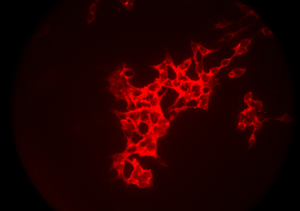
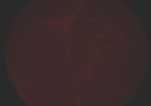
to damaged hippocampal area, we are exploring the use of appropriately
functionalized semiconductor quantum dots attached to neuron, glia,
astrocyte cells as biological labels. Through changes in the quantum
dot response as a function of the cell environment and interaction
with appropriately functionized silicon, silicondioxide and silicon
nitride surfaces, we expect to gain better understanding and control
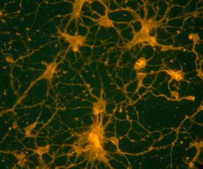
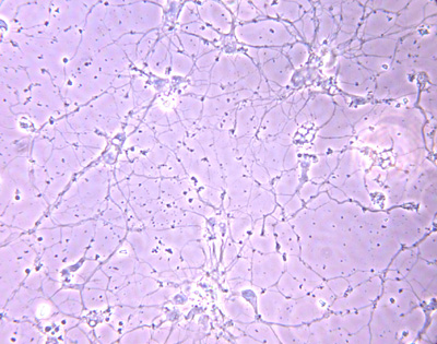
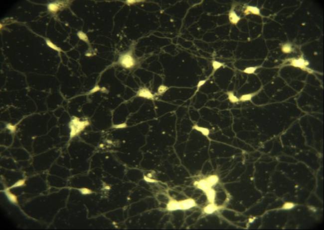
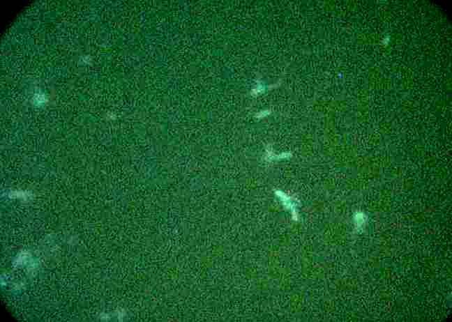
of the nature of the cell-surface interactions. Examples of such
use of semiconductor quantum dots is shown in the images below.
This is supported by the NSF Engineering Research Center for Biomimetic
Microelectronic Systems.
|